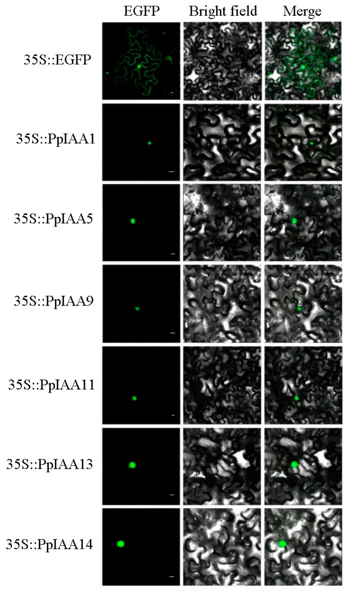
Figure 3.
Subcellular localization of selected peach Aux/IAA proteins. PpIAA1-GFP, PpIAA5-GFP, PpIAA9-GFP, PpIAA11-GFP, PpIAA13-GFP and PpIAA14-GFP fusion proteins were transiently expressed in tobacco leaves and their subcellular localization was determined by confocal microscopy. The green fluorescent ball represented the localization of the fusion protein to the nucleus, and the green fluorescent curve represented the localization of the protein to the cytomembrane. The scale bar indicates 15 μm.

