Figure 1.
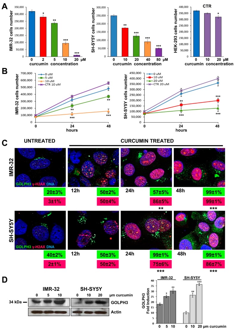
Curcumin provoked DNA damage in neuroblastoma cells and up-regulation of GOLPH3 with Golgi dispersal. (A) IMR-32, SH-SY5Y and non-tumorigenic control HEK-293 (CTR) cell lines were cultured in presence of various concentrations of curcumin for 24 h. (B) Cells were cultured with two curcumin concentrations for 24 and 48 h. At each harvest point, cells were trypsinized and counted in Trypan blue. Untreated cells (curcumin 0 µM) were cultured with 0.1% DMSO. Non-tumorigenic control HEK-293 cells (CTR) were cultured with the highest curcumin concentrations used for each NB cell line. Data are representative of three independent experiments ± SD. (C) Immunofluorescence analysis of IMR-32 and SH-SY5Y cells cultured with 10 or 20 µM curcumin respectively for 12, 24 and 48 h using anti-GOLPH3 (green) and anti-γH2AX (red). Cells were counterstained with DAPI to visualize nuclei (blue). Untreated cells were cultured with 0.1% DMSO. (Magnification 40×). In green and red boxes are reported the percentages of GOLPH3 and γH2AX positive cells respectively. Data are representative of three independent experiments ± SD. (D) IMR-32 and SH-SY5Y cells were cultured in presence of two concentrations of curcumin for 48 h, lysed, subjected to Western blot analysis and probed with anti-GOLPH3 antibody. Controls (curcumin 0 µM) were treated with 0.1% DMSO. Protein level was quantified by densitometry, normalized to the content of the loading control protein (actin) and visualized by histogram. Data are representative of three independent experiments ± SD (* p < 0.05; ** p < 0.01; *** p < 0.001)).

