Figure 3.
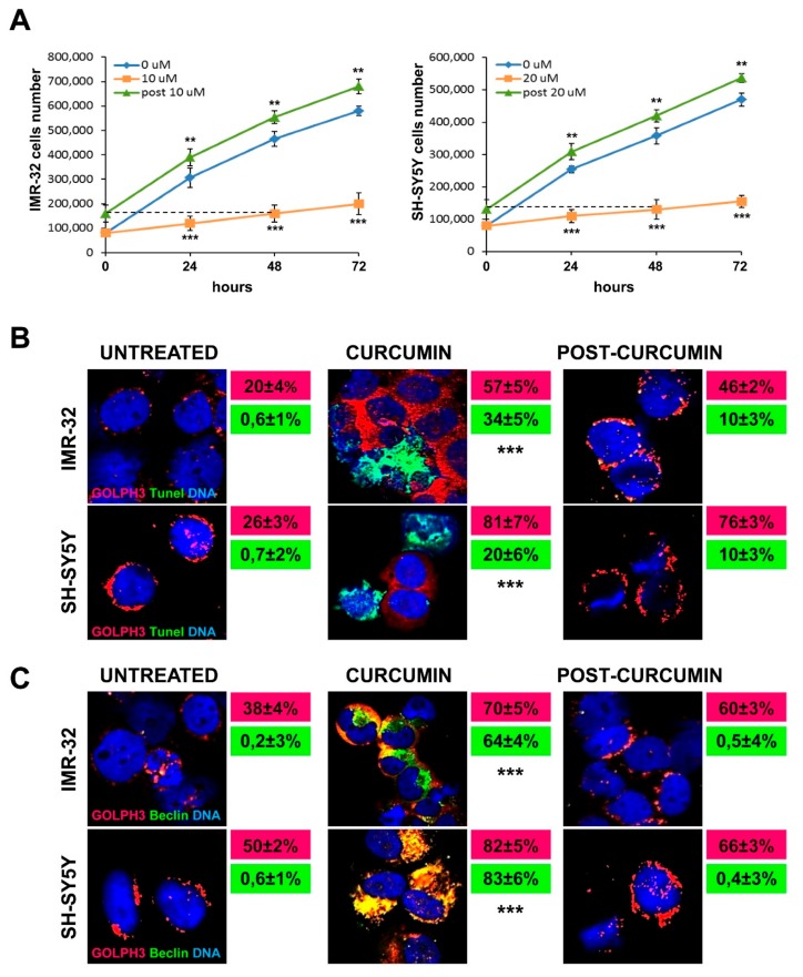
Golgi fragmentation is related to autophagy, not to apoptosis in neuroblastoma cells. (A) Comparison among the daily growth rate of untreated cells, of cells treated with curcumin and of cells recovering from 48 h of curcumin treatment, during three days of observation. For the recovery experiment (indicated as post 10 µM and post 20 µM) cells were incubated for 48 h with curcumin 10 or 20 µM and then washed and cultured in drug-free medium for 24, 48 and 72 h. The dashed line indicates the starting point of the recovery experiment observation, corresponding to the 48 h of curcumin treatment. At each harvest point, cells were trypsinized and counted in Trypan blue. Controls (curcumin 0 µM) were treated with 0.1% DMSO. Data are representative of three independent experiments ± SD. (B) Immunofluorescence analysis of IMR-32 and SH-SY5Y cells cultured with 10 or 20 µM curcumin respectively for 48 h and after 48 h post-Curcumin depletion, using anti-GOLPH3 (red) and TUNEL staining (green) to reveal chromatin condensation. In red and green boxes are reported the percentages of GOLPH3 and tunel positive cells respectively. (C) Immunofluorescence analysis of IMR-32 and SH-SY5Y cells using anti-GOLPH3 (red) and anti-Beclin-1 (green). In red and green boxes are reported the percentages of GOLPH3 and Beclin-1 positive cells respectively. Cells were counterstained with DAPI to visualize nuclei (blue). Untreated cells were cultured with 0.1% DMSO. (Magnification 40×). Data are representative of three independent experiments ± SD (** p < 0.01; *** p < 0.001).

