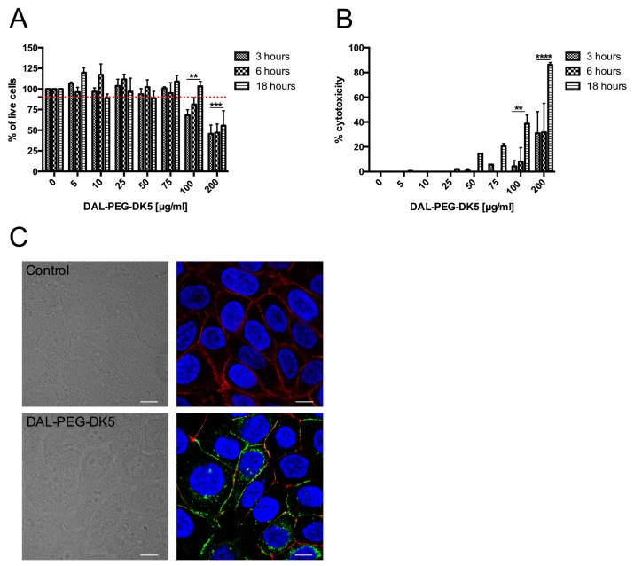
Figure 3.
The influence of DAL-PEG-DK5 on physiology of human keratinocytes. The potentially toxic effect of DAL-PEG-DK5 on HaCaT cells was evaluated using (A) MTT and (B) LDH assay. Cells were plated on 96-well plates and incubated overnight. Next, keratinocytes were treated with the peptide at different concentrations (5–200 μg/mL) for 3, 6, 18 h. Mean ± SD n = 2. ** p < 0.005 *** p < 0.001 **** p < 0.0001; 2way ANOVA. (C) Morphology of HaCaT cells was examined by confocal laser scanning microscopy. HaCaT cells were treated with CFS-conjugated DAL-PEG-DK5 (25 μg/mL) for 30 min at 37 °C and were stained with: DAPI and phalloidin for nuclear detection and actin cytoskeleton detection, respectively. Blue – DNA; red – f-actin; green-peptide conjugate; scale bar: 10 μm. Images present single slice of XY stacks.

