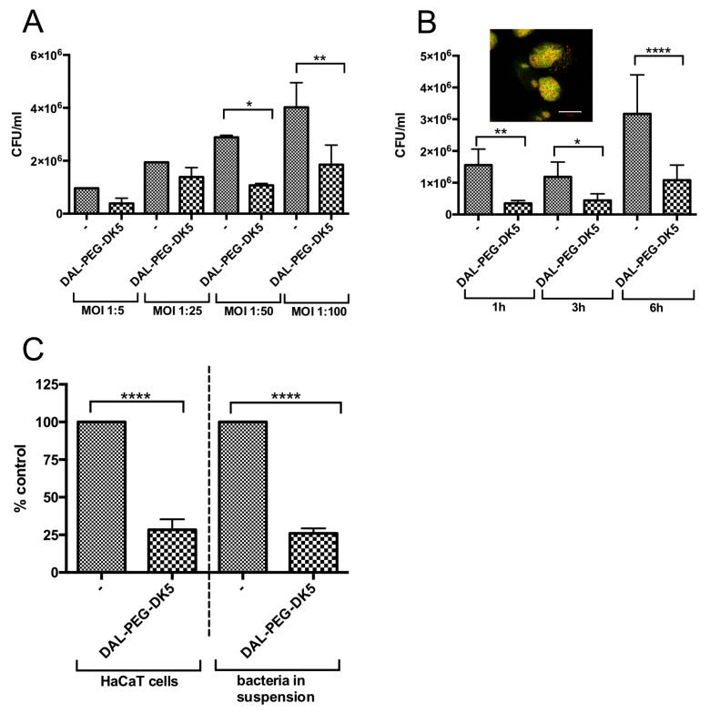
Figure 4.
Bactericidal activity of DAL-PEG-DK5 against intracellular S. aureus. (A) USA300 survival within infected HaCaT cells. Keratinocytes were infected with USA300 (MOI 1:5, 1:25, 1:50, 1:100) for 2.5 h and treated with DAL-PEG-DK5 (50 μg/mL) for 3 h. Afterwards, keratinocytes were lysed and plated on agar plates for counting of bacteria. The number of viable bacterial cells is expressed as CFU/mL with respect to the number of intracellular bacteria in the corresponding control samples. The data shown is representative of two separate experiments performed in triplicate. Mean ± SD n = 2. * p < 0.01; ** p < 0.005; one-way ANOVA. (B) Keratinocytes were infected with USA300 (MOI 1:50) for 2.5 h and treated with DAL-PEG-DK5 (50 μg/mL) for indicated time points (1, 3, 6 h). Afterwards, keratinocytes were lysed and plated on agar plates for counting of bacteria. The number of viable bacterial cells is expressed as a CFU/mL with respect to the number of intracellular bacteria in the corresponding control samples. Mean ± SD n = 2. * p < 0.01; ** p < 0.005; **** p < 0.0001; one-way ANOVA. For confocal laser scanning microscopy HaCaT cells were infected with USA300 (MOI 1:50) for 2.5 h, then treated with DAL-PEG-DK5 (50 μg/mL) for 3 h. Afterwards cells were stained with SYTO 9 and PI. Viable S. aureus cells are stained green while red signals represent dead bacteria and the host cell’s nuclear DNA stained with SYTO 9 and PI. Scale bar: 10 μm. Image presents single slice of XY stacks. (C) Keratinocytes were infected with USA300 (MOI 1:50) for 2.5 h and treated with DAL-PEG-DK5 (50 μg/mL) for 1 h. Afterwards, keratinocytes were lysed and plated on agar plates for counting of bacteria. In parallel, MRSA (2 × 10⁶ CFU/mL) were incubated with DAL-PEG-DK5 (50 μg/mL) for 1 h and then plated on agar plates. The number of viable bacterial cells is expressed as a % of control with respect to the number of intracellular bacteria/bacteria in suspension in the corresponding control samples. Mean ± SD n=2. **** p < 0.0001; one-way ANOVA.

