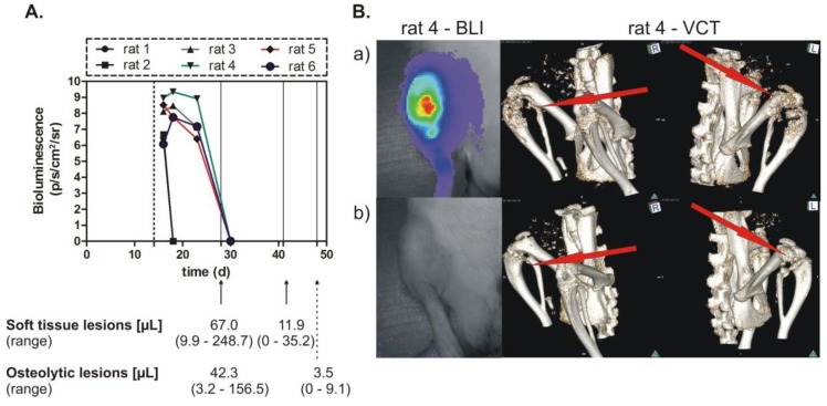
Figure 4.
Inhibition of breast cancer skeletal metastasis upon conditional miRNA-mediated OPN knockdown. (A) Bioluminescence imaging (BLI) detection of tumor cells and measurement of the volume of soft tissue and osteolytic lesions. Cells of the clone O1 were inoculated into nude rats, which received doxycycline for 2 weeks (dashed line indicates the end of this administration) and were exposed to miRNA treatment thereafter. The time after tumor cell inoculation (days) is given on the x-axis; bioluminescence was measured in photons/second/cm2/steradian; the volume of soft tissue and osteolytic lesion is indicated at 28, 42 or 49 days after tumor cell inoculation or 14, 28, or 35 days of miRNA treatment. (B) BLI and volume computed tomography (VCT) images of tumors and skeletal metastasis. In the upper panel (a) are the BLI image (left) and VCT scans (middle and right), which show the status at 19 and 28 days after tumor cell inoculation, respectively; the lower panel (b) shows the BLI image (left) and VCT scans (middle and right), which indicate the status at 30 and 49 days after tumor cell inoculation, respectively.

