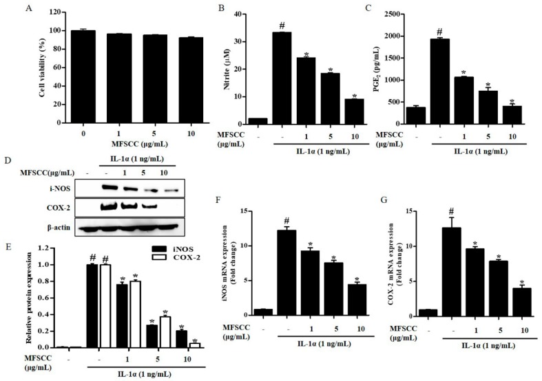
Figure 1.
Effect of MFSCC on cell viability and inflammation in rat primary chondrocytes. (A) The cytotoxic effects of MFSCC were examined by CCK-8 assay and cell cultures were treated with different concentrations of MFSCC (0, 1, 5, and 10 μg/mL) for 24 h. The MFSCC showed no cytotoxicity in rat primary chondrocytes. The results are indicated as mean ± SEM (* p < 0.05 compared with the control group). (B,C) Anti-inflammatory equities of MFSCC (0, 1, 5, and 10 μg/mL) were determined by the estimation of NO and PGE2 production. (D–G) The expression of iNOS and COX-2 was detected by western blot and qRT-PCR. Rat primary chondrocytes were co-treated with different concentrations of MFSCC (0, 1, 5, and 10 μg/mL) and 1 ng/mL IL-1α for 24 h. The results are expressed as mean ± SEM (# p < 0.05 compared with the control group; * p < 0.05 compared with the IL-1α group).

