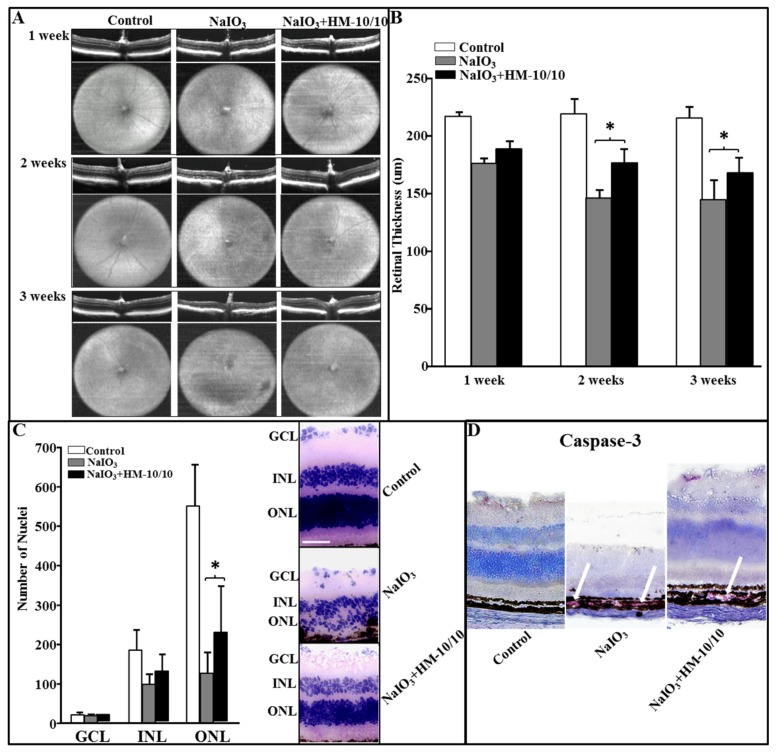
Figure 4.
HM-10/10 treatment prevents degenerative changes in retina induced by intravenous NaIO3. Eye in vivo optical coherence tomography (OCT) images and the fundus photographs were taken after one, two, and three weeks of NaIO3 treatment as described under Materials and Methods. The mice were sacrificed at the end of three weeks. Retinal sections were scanned. (A) Degenerative changes shown by representative OCT and fundus images from Control, NaIO3, and NaIO3 + HM-10/10 groups of mice. Horizontal and vertical extents of fundus images are 1.4 × 1.4 mm, respectively. (B) Retinal thickness measurements from all three weeks of the study represented as bar graphs. (C) The numbers of nuclei were counted from each layer as described under Materials and Methods. The representative histopathology images showing decreased number of nuclei with NaIO3 treatment as compared to control and partial recovery in the number of nuclei with HM-10/10 treatment to that of control. Bar equals 50 μm. GCL: Ganglion cell layer; ONL: Outer nuclear layer; INL: Inner nuclear layer. (D) Immunohistochemistry staining of cleaved Caspase-3 (red) of retinal tissue sections. Bonferroni method was used to adjust the p < 0.0016 for multiple pairwise comparisons.

