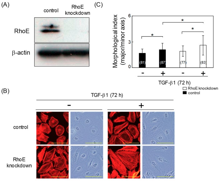
Figure 2.
RhoE knockdown increases the degree of cell elongation in HeLa cells treated with TGF-β1. (A) Knockdown efficiency of RhoE in HeLa cells. HeLa cells were transfected with siRhoE-A. Luciferase small interfering RNA (siRNA) was used as a control and β-actin expression was used as a loading control. (B) Morphological changes in HeLa cells transfected with siRhoE-A. Cells were treated with 1 ng/mL TGF-β1 for 72 h. F-actin was visualized using tetramethylrhodamine isothiocyanate (TRITC)-conjugated phalloidin. Scale bars represent 100 μm. (C) Quantitative analysis of cell morphology of HeLa cells in (B). The lengths of the major and minor cell axes were measured using NIH-Image software. The ratios of the major to minor axes of cells were used to determine the degree of cell elongation. More than 77 cells were measured per condition for each experiment. Numbers used for the experiments are shown in parentheses. Data in each column represent the mean and standard deviation. Statistical significance was assessed using one-way ANOVA with a post hoc Tukey–Kramer HSD test. *p < 0.01. Similar results were obtained in two independent experiments.

