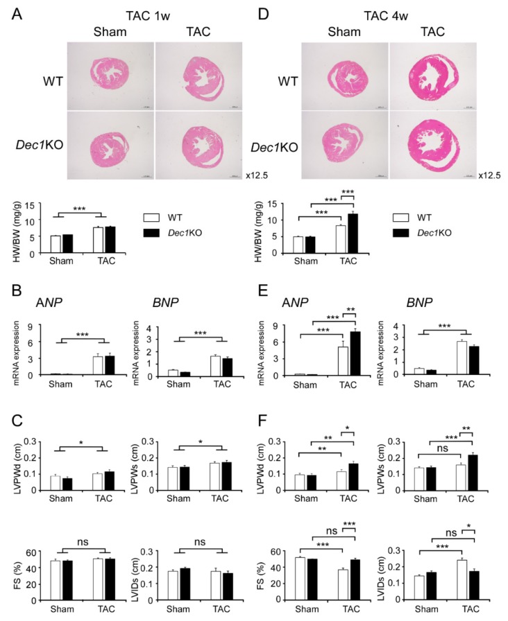
Figure 1.
Differentiated embryonic chondrocyte gene 1 (Dec1) deficiency protects cardiac function against transverse aortic constriction (TAC)-induced pressure overload. (A) Assessment of cardiac phenotype in wild-type (WT) and Dec1 knock-out (Dec1KO) mice at one week (1w) after TAC (n = 11–13 mice per group) and sham treatment (n = 5–6 per group). Hematoxylin and eosin (H&E) staining of WT and Dec1KO hearts, magnification 12.5×. Heart weight/body weight (HW/BW). White box indicates WT and black box indicates Dec1KO. (B) The relative messenger RNA (mRNA) expression of the hypertrophy markers, ANP (atrial natriuretic peptide) and BNP (B-type natriuretic peptide), in WT and Dec1KO mice at one week after TAC and sham. (C) Echocardiographic analysis of WT and Dec1KO mice at one week after TAC and sham. FS: fractional shortening; LVIDs: left ventricular internal dimension at the end of systole; LVPWd and LVPWs: left ventricular posterior wall thickness at the end of diastole and at the end of systole, respectively. (D) Assessment of cardiac phenotype in WT and Dec1KO mice at four weeks (4w) after TAC and sham treatment (n = 8–11 per group). (E) The relative mRNA expression of the hypertrophy markers, ANP and BNP, in WT and Dec1KO mice at four weeks after TAC and sham treatment. (F) Echocardiographic analysis of WT and Dec1KO mice at four weeks after TAC and sham treatment. Multiple comparisons between sham and TAC groups were analyzed by two-way ANOVA with Tukey–Kramer post hoc test. Data are the means ± standard error of the mean (SEM). * p < 0.05, ** p < 0.01, *** p < 0.001; NS: not significant.

