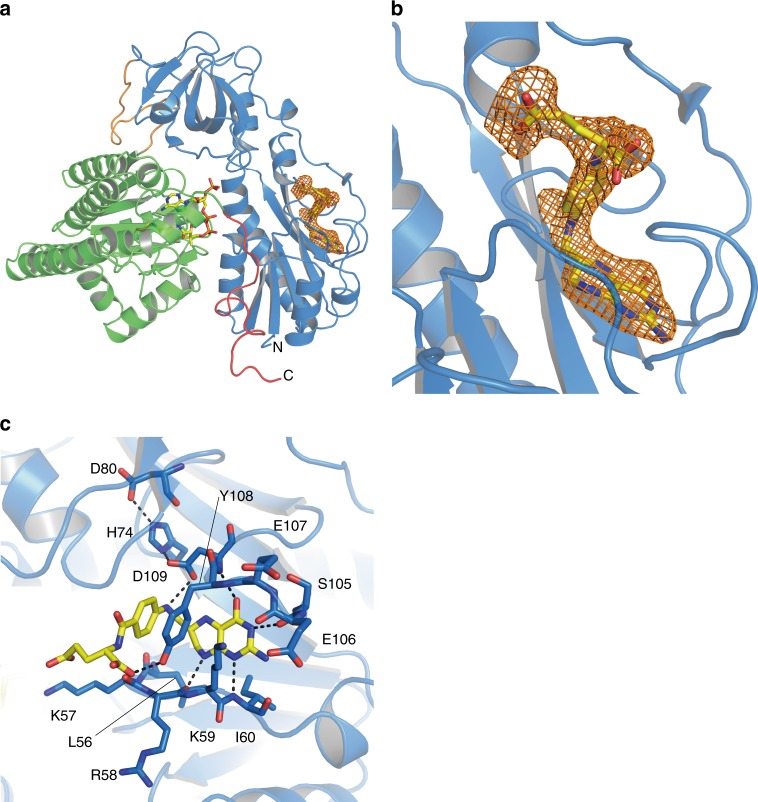
Fig. 3.
Structure of THF-bound HypX (PDB ID: 6J1F). a Electron density map (Fo-Fc polder omit map) contoured at 3σ for THF in wild-type HypX, which is shown with an orange mesh. b Close-up view of the THF binding region. c Interactions between THF and HypX. Pterin ring of THF is sandwiched by the β3-α3 (residues 53–62) and the β5–β6 (residues 103–114) loops. His74, Asp80, and Asp109 form the hydrogen bonding network to fix the orientation of Asp109. Hydrogen bonds are shown in dashed lines

