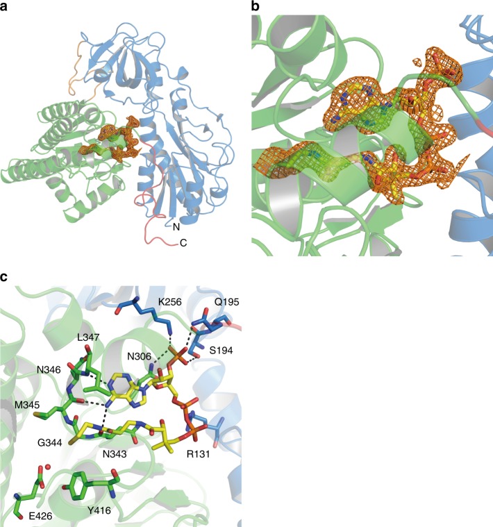
Fig. 4.
a Electron density map (Fo-Fc polder omit map) contoured at 3σ for CoA in wild-type HypX, which is shown with an orange mesh. b Close-up view of the CoA binding region. c Interactions between CoA and HypX. CoA and several amino acid residues are shown in the stick model. A red ball stands for the oxygen atom of a water molecule. CoA interacts with amino acids in the both of the N-terminal domain (blue) and the C-terminal domain (light green). Hydrogen bonds are shown in dashed lines

