Fig. 5.
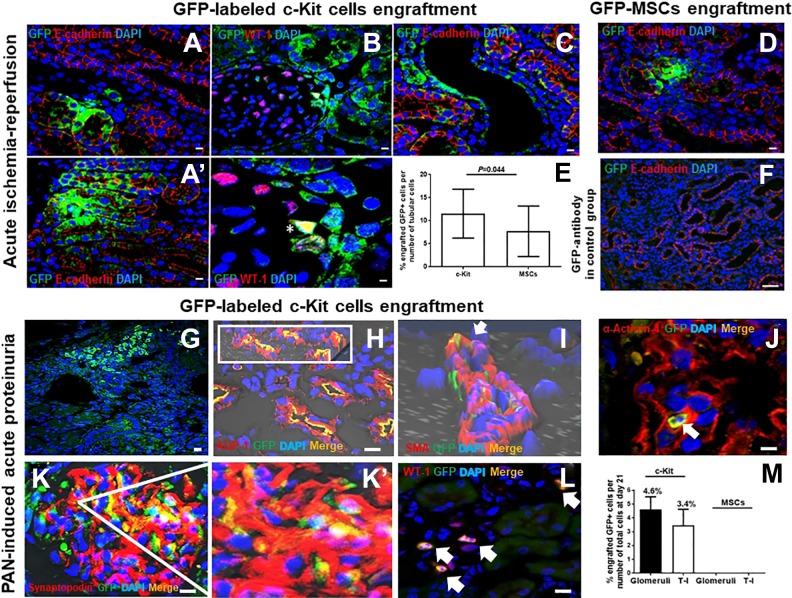
Progenitor/stem cell engraftment within the kidneys after suprarenal aorta delivery. In the acute ischemia-reperfusion model, GFP-labeled c-Kit progenitor/stem cells exhibited multi-compartment engraftment, including tubules, as shown by E-cadherin co-staining (A, A’); glomeruli in Bowman’s capsule and podocyte (B), as shown by WT-1 co-staining (*); and vascular (C). GFP-MSCs engrafted within the kidneys less frequently when compared with GFP-labeled c-Kit progenitor/stem cells (D, E). GFP antibody was used in the control group (F). Adapted from the method of Rangel and colleagues13. In the acute proteinuria model induced by puromycin aminonucleoside (PAN), GFP-labeled c-Kit progenitor/stem cells also engrafted into multi-compartments of the kidneys (G). Cells in (F) co-stained for aquaporin-1 (AQP1) (H; insert shows 3D confocal image), smooth muscle actin (SMA) (arrow; 3D confocal image) (I), α-Actinin-4 (arrow) (J), synaptopodin (K, K’), and WT-1 (arrows) (L). GFP-labeled c-Kit progenitor/stem cells engrafted into tubules and glomeruli in higher numbers when compared with GFP-MSCs (M). Adapted from the method of Rangel and colleagues14. Scale bars represent 20 µm for confocal images.

