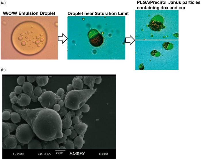
Figure 12.
Optical microscope pictures depicting PLGA-Precirol Janus particles in the presence DOX and CUR. Compartmentalization of the drug particles is demonstrated visually in the optical microscope pictures through localization of the red DOX to the Precirol® compartment and bright yellow CUR in the PLGA compartment, as it can be clearly observed in the optical microscope pictures in Figure 12(a). (a) PLGA/Precirol Janus particle formation from W/O/W emulsions. Step 1: W/O/W emulsion consisting of W/O emulsion droplets containing DOX inside an O/W emulsion droplet containing CUR. Step 2: Initial phase separation of the polymer and lipid phases during solvent evaporation. The red color of the drug DOX can be clearly seen in the Precirol compartment (conical part of the ice cream cone), and the yellow color of CUR can be seen in the PLGA phase. Step 3: Formation of PLGA/Precirol particles upon complete solvent evaporation (b) Fully formed PLGA/Precirol particles in the absence of any drugs. (A color version of this figure is available in the online journal.)

