Figure 4.
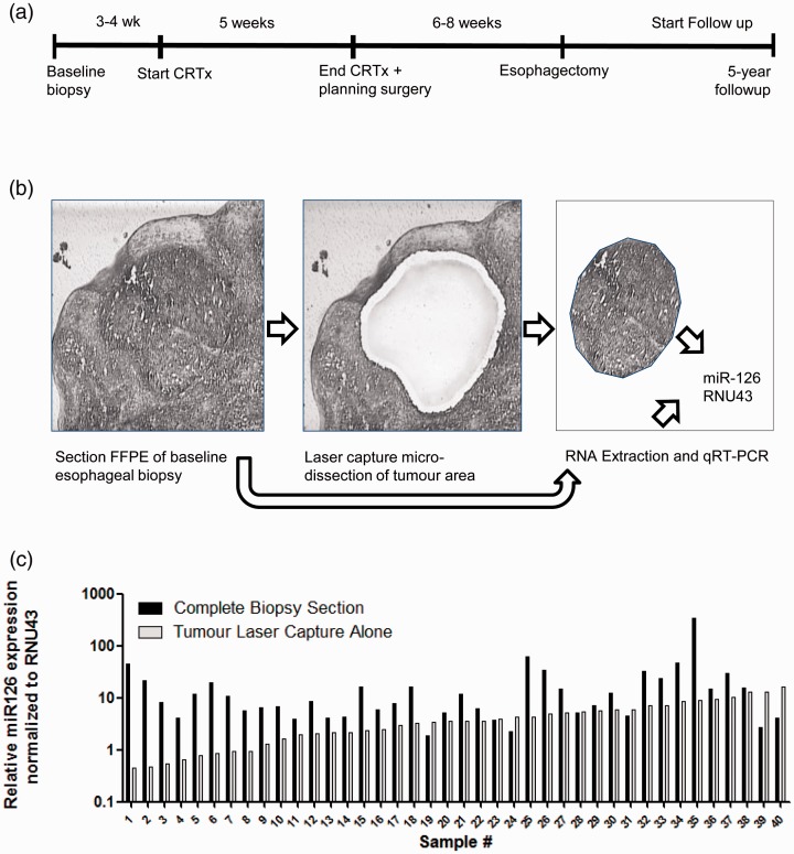
Evaluation of miR-126 levels in esophageal biopsies of patients with OAC. (a) The time-line of treatment of patients with OAC and their follow-up are shown. In total, 58 patients were treated with combined neoadjuvant chemoradiotherapy and surgical resection and patient overall survival was monitored for five years post treatment. (b) Esophageal biopsies taken at baseline containing both tumor and non-tumorous tissue including the squamous epithelium. Shown is a representative tissue section with HE-staining within the middle a tumor-island (Left panel). Tumor-specific RNA was obtained by laser capture microdissection microscopy (Middle panel). Following microdissection, the tumor-piece was captured in a small tube ready for RNA isolation. Also, RNA was extracted from the complete tissue section (Right panel). (c) Shown is the relative level of miR-126 normalized to RNU43 of 40 patients. Levels in tumor showed a wide variation (ranging from 0.4 to 16.4 normalized miR-126 values).

