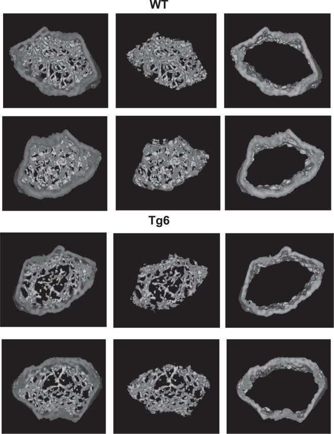
Figure 8.
Bone phenotype of 6–8 weeks old female Tg6 mice. The bone phenotype of 6–8 weeks old female Tg6 mice and their wildtype littermates was examined by μCT. Representative cross-sections microCT images of distal femora of WT versus Tg6 are shown. Dark grey area indicates mineralized bone. Data are means ± s.e.m.; n = 5–9 for each group of mice. Significance was determined by unpaired t test and indicated as *p < 0.05 and ***p < 0.001.

