Figure 1.
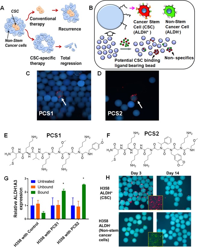
Identification of ALDH+ CSC-binding Peptoids. (A) Conventional therapies may eliminate the majority of cancer cells, but drug resistant subpopulations such as CSCs may remain intact, which can regenerate the tumor. Therapies directed towards eliminating CSCs could support complete elimination of the tumor. (B) Outline of the on-bead two-color (OBTC) Assay. H358 cells were sorted into ALDH+ (CSCs: stained red) and ALDH− (non-CSCs: stained green), were mixed 1:1 and exposed to bead (blue) library carrying peptoids on one-bead one-compound format. A bead binding only red cells indicates that the peptoid displayed on that bead recognizes a molecule only (or predominantly) found on CSCs, which is absent (or less abundant) on remaining cancer cells. (C,D) Beads (blue) bound only by red cells were picked as carrying highly CSC specific ‘hit’ peptoids, PCS1 and PCS2. (E,F) The structures of ‘hit’ peptoids PCS1 and PCS2, respectively. (G) Relative expression of ALDH1A3 was higher in PCS1 and PCS2 coated magnetic bead bound fractions than unbound or untreated H358. Error bars represent standard deviation between triplicates. *Represents p value < 0.05. (H) PCS2 displayed on tentagel beads preferentially recognize ALDH+ cells. PCS2-carrying tentagel beads bind to sorted and red Qdot stained ALDH+ cells at the day 3 (top row, left panel), but not to green Qdot stained ALDH− (bottom row, left panel). This binding ability of ALDH+ cell group was greatly reduced after 2 weeks (top row, right panel). Insert boxes are the red stained ALDH+ and green stained ALDH− cells. See also Supplementary Figs. S1–S2, and Table S1.

