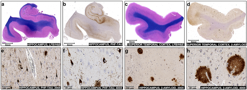
Fig. 1.
Pathologic NFT inclusions and amyloid-β plaques accumulate in vulnerable brain regions during Alzheimer’s disease. Sections of mid hippocampus were a stained with hematoxylin and eosin plus luxol fast blue (H&E/LFB) or b used for immunohistochemical staining with PHF-tau antibody. Sections of the superior temporal gyrus were c stained with H&E/LFB or d used for immunohistochemical staining with amyloid-β antibody. Neurofibrillary degeneration and scattered neuritic plaques are visualized at × 200 (e) and at × 400 (f) in the hippocampal section incubated with PHF-tau antibody. Diffuse and dense core plaques are visualized at × 200 (g) and at × 400 (h) in the superior temporal gyrus section incubated with amyloid-β Antibody

