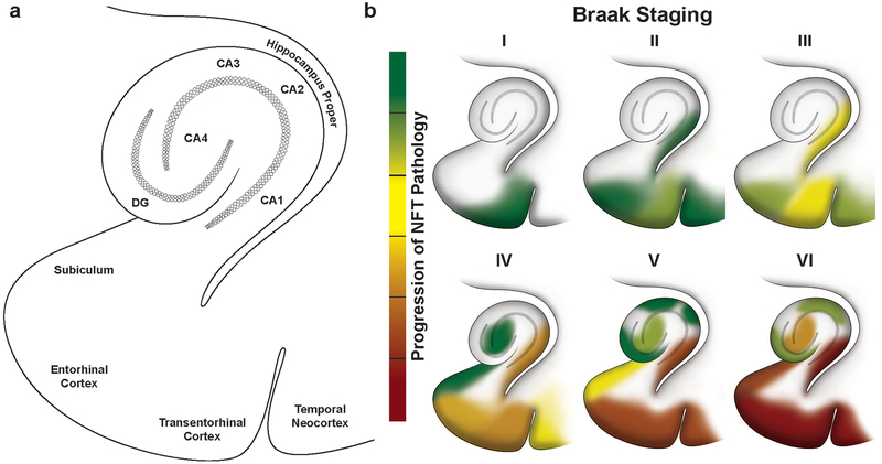
Fig. 2.
Progressive accumulation of NFTs within the transentorhinal and entorhinal cortex and hippocampal regions along Braak stages of neurofibrillary degeneration in Alzheimer’s disease, a Sub-regions within the transentorhinal and entorhinal cortex and hippocampus that are differentially vulnerable to developing NFTs and succumbing to disease, b The progression of NFT appearance and accumulation during Braak staging of AD from stage I to VI. Green color denotes low abundance of NFTs and red denotes high abundance of NFTs within a sub-region. Note that the CA4 sub-region is also referred to as the “hilus”

