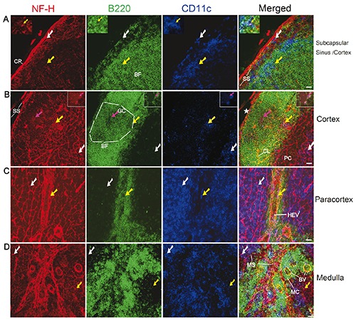
Figure 2.

Distribution of nerve fibres in the subcapsular sinus (A), cortex (A-B), paracortex (C), and medulla (D) in the mesenteric lymph node of a C57BL/6 mouse. Antibodies against NF-H (red), B220 (green), and CD11c (blue) label mainly nerve fibres, B cells, and DCs, respectively. The white arrows show B220-CD11c+ DCs that have a close association with the nerve fibres. The yellow arrows indicate B220+CD11c+ DCs that have a close association with the nerve fibres. A) The inserted windows show B220+CD11c+ DC (also indicated by yellow arrows) that has a close association with the nerve fibres. B) The magenta arrows (also in inserted windows) indicate a few B cells (B220+CD11c-) that have a close association with the nerve endings (appearing as red dots around B cells). The star indicates a torn site of the subcapsular sinus. The white dashed lines outline the germinal centre (identified from its morphology). All images are generated by using maximum intensity projection of 13 optical slices. Stack size: 6.0 μm; optical slice interval: 0.5 μm. CP, capsule; BF, B cell follicle; GC, germinal centre; SS, subcapsular sinus; CL, capillary; HEV, high endothelial venules; PC, paracortex; MS, medullary sinus; MC, medullary cord; BV,: blood vessel.; Objective lens: 40x; scale bar: 20 μm.
