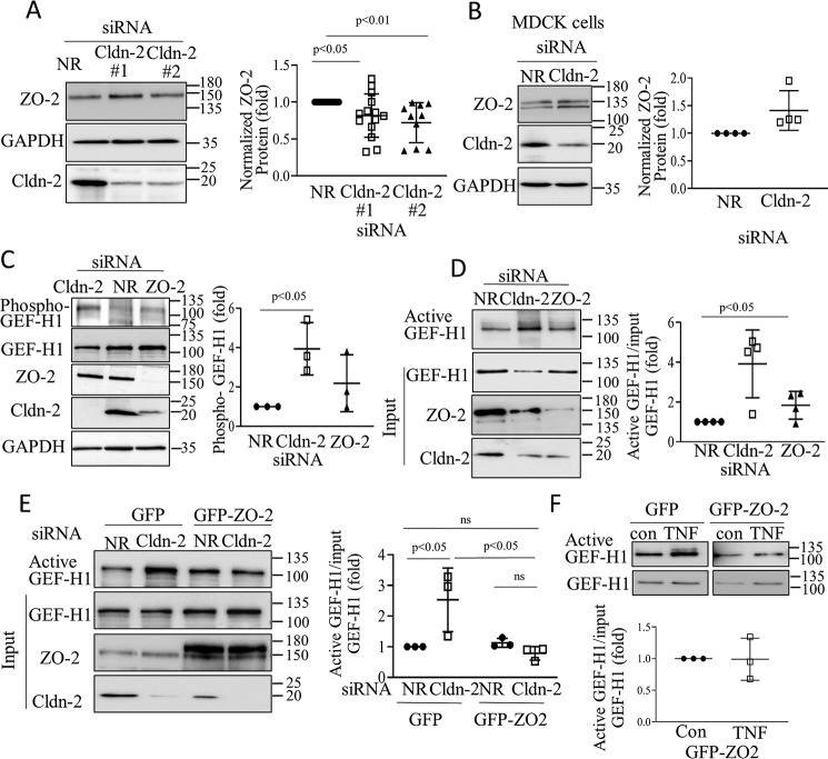
Figure 5.
A and B, LLC-PK1 (A) or MDCK (B) cells were transfected with NR or Cldn-2 siRNA, and 48 h later the indicated proteins were detected by Western blotting. The corresponding graphs show ZO-2 normalized with GAPDH (fold from NR siRNA transfected control) (means ± S.D.; in A, n = 14 and 12 for the two siRNAs, respectively; in B, n = 4). C and D, LLC-PK1 cells were transfected with NR or Cldn-2 or ZO-2 siRNA as indicated. In C, phospho-GEF-H1 was detected and quantified using an antibody against pSer-885 GEF-H1. The graph shows the phospho–GEF-H1 signal normalized to total GEF-H1 and expressed as fold over control. In D, 48 h after transfection, active GEF-H1 was detected and quantified as earlier (means ± S.D., n = 3). The indicated proteins were also detected in total cell lysates. E and F, LLC-PK1 cells expressing GFP–ZO-2 or the eGFP control vector were transfected with NR or Cldn-2 siRNA (E) or treated with 10 ng/ml TNFα for 10 min (F). Active GEF-H1 was detected and quantified as earlier (means ± S.D., n = 3). The indicated proteins were also detected in total cell lysates.

