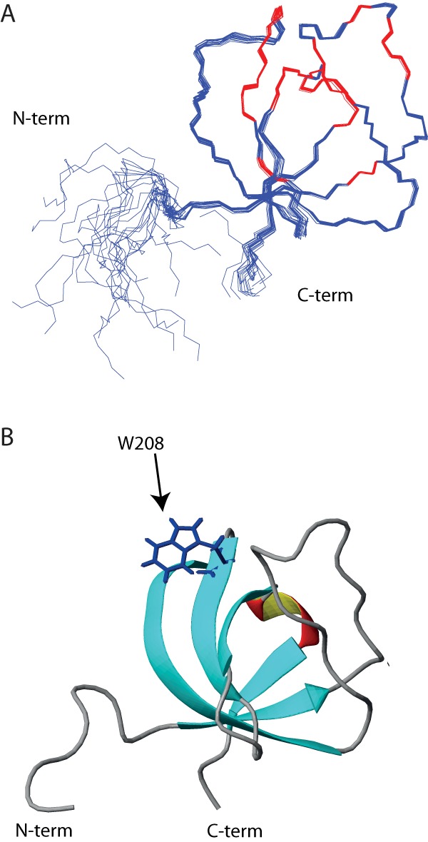
Figure 3.

The human ITK SH3 domain solution structure. A, backbone trace of the 20 structures comprising the lowest-energy NMR ensemble is shown. Red color indicates residues affected by ligand binding. B, ribbon representation of one of the ITK SH3 domain structures shown in A. The tryptophan at position 208, which is critical for polyproline ligand binding, is indicated.
