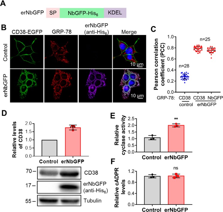
Figure 6.
CD38, retained in the ER, could not elevate cellular cADPR levels. A, schematic of erNbGFP, the ER-retained NbGFP with a His6 tag. SP, signal peptide of calreticulin; KEDL, an ER retention signal motif. B, CD38-EGFP/HEK293 cells were infected with a lentivirus encoding erNbGFP. The resulting cell line was stained with anti-GRP78, an ER marker, anti-His6 for erNbGFP, and DAPI. The fluorescence developed by antibody and dyes, together with that from the EGFP tag of CD38, was imaged under a Nikon A1 confocal microscope. Green, CD38; red, GRP78; purple, erNbGFP; blue, DAPI for the nucleus. C, PCCs were calculated by overlapping the signals of CD38 or erNbGFP with those of GRP78 from the experiment in B. D, the total expression levels of CD38 were analyzed by Western blotting, together with erNbGFP, blotted by anti-CD38 and anti-His6, respectively. E, the ADP-ribosyl cyclase activities of the lysates were measured with an NGD assay. F, the cellular cADPR levels were measured by a cycling assay. All experiments were repeated at least three times (mean ± SD; n = 3; Student's t test; **, p < 0.01, 0.0001; ns, not significant).

