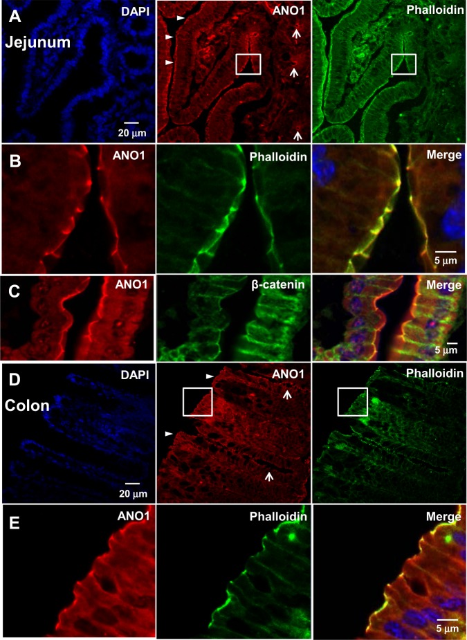
Fig. 3. ANO1 is expressed in the apical membranes of the small intestine and colon.
a–e Immunofluorescence images of the jejunum (a–c) and colon (d, e) stained with antibodies specific for ANO1, as well as phalloidin and β-catenin, markers for apical and lateral membranes, respectively. Arrowheads indicate the strong expression of ANO1 in the apical membrane of villi and surface epithelial cells in the jejunum (a) and colon (d). ANO1 showed relatively weak expression in the apical membrane of the crypt in the jejunum and colon (arrows) compared to the villi and surface region (arrowheads). b, c, e Magnified images of epithelial cells in the square regions of (a) and (d). ANO1 was largely colocalized with phalloidin at the apical membrane (b) but was not colocalized with β-catenin, which is the lateral membrane marker (c)

