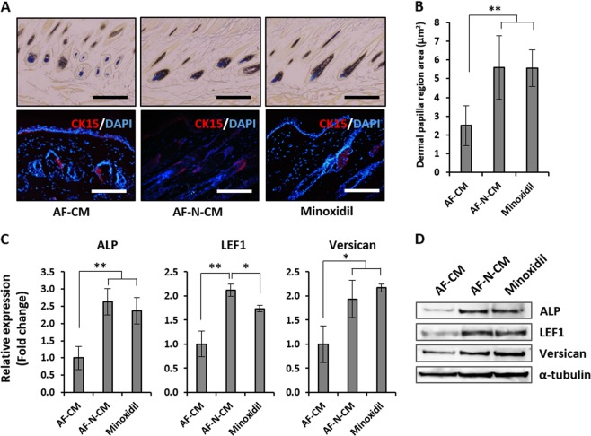
Fig. 6. Histological analysis of amniotic fluid–derived mesenchymal stem cell overexpressing Nanog (AF-N-MSC)–derived conditioned media (AF-N-CM)-treated dorsal skin.
a Expression patterns of the structural indicators at 5 days post-treatment: AP for hair follicle dermal papilla cell population (top) and CK15 for bulge stem cells (bottom). Quantification of the AP-positive dermal papilla region (right). Scale bar, 200 µm. b Quantification of the hair follicle dermal papilla region area (μm2) determined by ImageJ. c Expression of hair growth markers (ALP, LEF1, and Versican) in AF-CM, AF-N-CM, and minoxidil treatments, as determined by quantitative real-time reverse transcriptase–polymerase chain reaction. d Protein levels of ALP, LEF1, and Versican in AF-CM, AF-N-CM, and minoxidil treatments, as determined by western blot. Data are represented as the mean ± SD (n = 3). *p < 0.05, **p < 0.01

