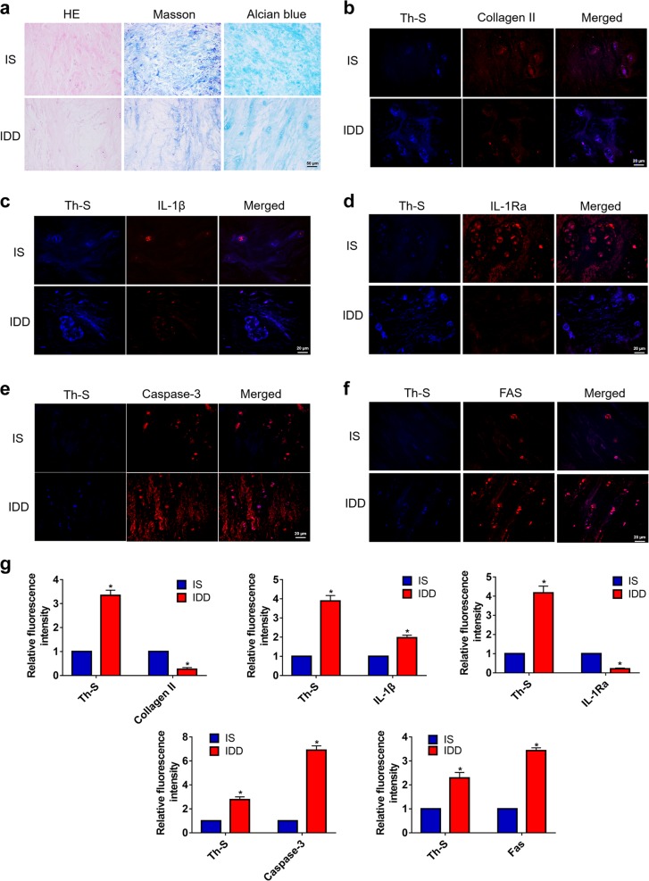
Fig. 1. The aggregation of hIAPP in human NP tissues.
a Representative images of histological staining of NP tissues from patients with idiopathic scoliosis (IS) or degenerative disc disease (IDD). (b-f) Immunofluorescence staining for collagen II b, IL-1β c, IL-1Ra d caspase-3 e, and FAS f and costaining with Th-S. Th-S staining showed the amyloidosis of hIAPP. g The quantitative analysis of the relative fluorescence intensity of Th-S staining and collagen II, IL-1β, IL-1Ra, caspase-3, and FAS immunofluorescence using Image-Pro Plus 6.0. The data are presented as the mean ± SD (n = 5). *P < 0.05 vs. the corresponding IS group

