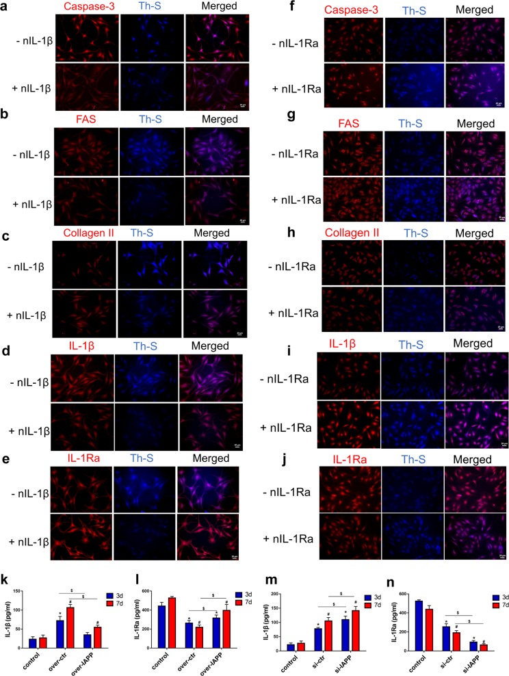
Fig. 7. IL-1β/IL-1Ra signaling regulated ECM metabolism and cell apoptosis through the deposition of hIAPP aggregates in human NP cells.
a–e Representative images of immunofluorescence for caspase-3 a, FAS b, collagen II c, IL-1β d, and IL-1Ra e and costaining with Th-S. hIAPP was overexpressed in human NP cells with or without nIL-1β (1 μg/mL) under static compression for one week. f–j Representative images of immunofluorescence for caspase-3 f, FAS g, collagen II h, IL-1β i and IL-1Ra j and costaining with Th-S. hIAPP was knocked down in human NP cells with or without nIL-1Ra (1 μg/mL) under static compression for one week. k–n The content of IL-1β and IL-1Ra in the culture supernatant was measured by ELISA kits in hIAPP-overexpressed k, l or hIAPP-silenced m, n NP cells under compression treatment for 3 d or 7 d. The data are presented as the mean ± SD (n = 3). *P < 0.05 vs. the 3-d control group. #P < 0.05 vs. the 7-d control group. $P < 0.05 vs. the corresponding group

