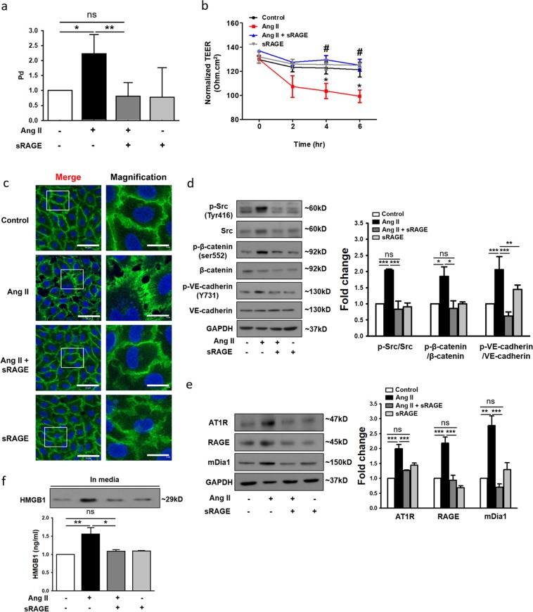
Fig. 4. Soluble RAGE (sRAGE) attenuates Ang II-induced endothelial hyperpermeability.
a HUVECs were treated with sRAGE (2 μg/ml) for 1 h and were then subjected to Ang II treatment for 4 h. Endothelial permeability was assessed in media from the lower chambers after the addition of FITC-dextran 40 to the upper chambers for 1 h (n = 3 for each lane). b TEER was measured every 2 h. *p < 0.05 vs. control, #p < 0.05 vs. Ang II (n = 3 for each lane). c Immunocytochemistry of VE-cadherin (green) and DAPI (nuclei, blue) was performed, and samples were directly examined under a confocal microscope (white scale bars: 50 μm in merged images and 5 μm in magnified images; ×400 magnification). The main images were selected from representative regions. d HUVEC extracts were analyzed by western blotting to assess phospho-Src, phospho-β-catenin, and phospho-VE-cadherin expression. Expression values were normalized to those of Src, β-catenin, and VE-cadherin (n = 4 for each lane). e The protein levels of AT1R, RAGE, and mDia1 were determined by western blotting. Expression was normalized to that of GAPDH (n = 3 for each lane). f HMGB1 release in supernatants was measured by ELISA and western blotting (n = 3 for each lane). The values are presented as the means ± SEMs. *p < 0.05; **p < 0.01; ***p < 0.001; ns not significant by one-way analysis of variance (ANOVA) followed by Tukey’s multiple comparisons test

