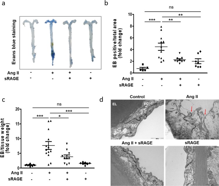
Fig. 5. Effects of sRAGE on Ang II-induced endothelial hyperpermeability in vivo.
Twelve-week-old ApoE KO mice were used to investigate endothelial hyperpermeability. Ang II was injected into mice with or without sRAGE for 6 weeks. Then, Evans blue (EB) dye in saline was administered via the jugular vein. a Representative photographs of aortas stained with Evans blue in ApoE KO mice. b Quantification of positive areas of Evans blue staining in the aortas, as estimated using ImageJ. c Evans blue dye was eluted from the aortas by incubation with formamide. The amount of dye was quantified by spectrophotometry at 610 nm. d Representative TEM images of the EC junction area (original magnification, ×30,000). EL elastic lamina, EC endothelial cell; scale bar, 1000 nm. Control group, n = 10; Ang II group, n = 13; Ang II + sRAGE group, n = 11; sRAGE group, n = 7. The results are representative of at least four separate experiments. The values are presented as the means ± SEMs. *p < 0.05; **p < 0.01; ***p < 0.001; ns not significant by one-way analysis of variance (ANOVA) followed by Tukey’s multiple comparison test

