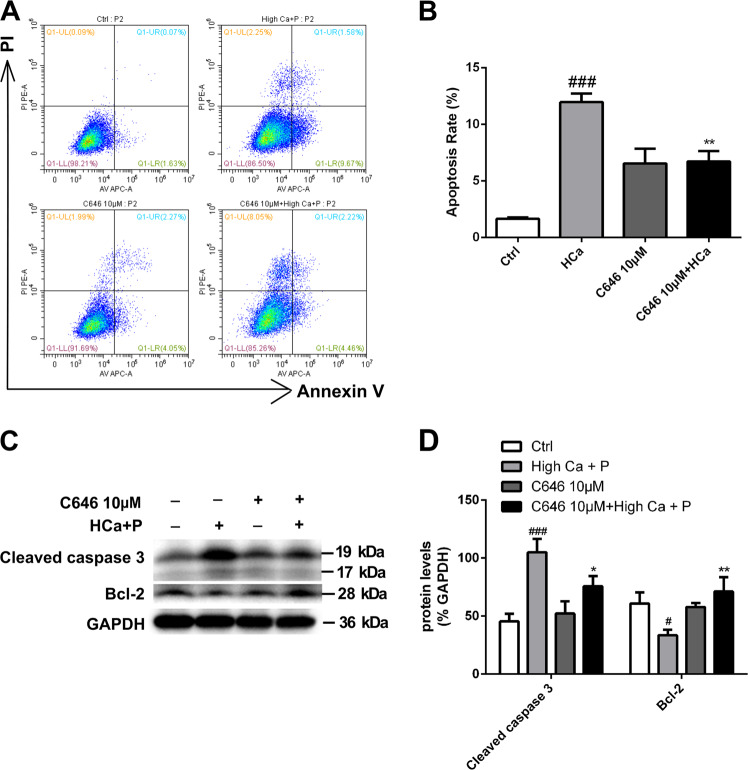
Fig. 3. The effect of C646 on apoptosis and proliferation of pAVICs after calcification.
a, b Flow cytometry analysis of apoptosis revealed that C646 inhibited apoptosis induced by high-calcium/high-phosphate treatment. The right bar graph shows the results of statistical analysis of the apoptosis ratios. c, d The expression of cleaved caspase 3 and Bcl-2 was assessed by western blotting. The relative protein levels were normalized to GAPDH levels. The data are shown as the means ± standard errors of the means of triplicates and are representative of three independent experiments performed (#p < 0.05, ###p < 0.001 compared with control; *p < 0.05, **p < 0.01 compared with high Ca+P)

