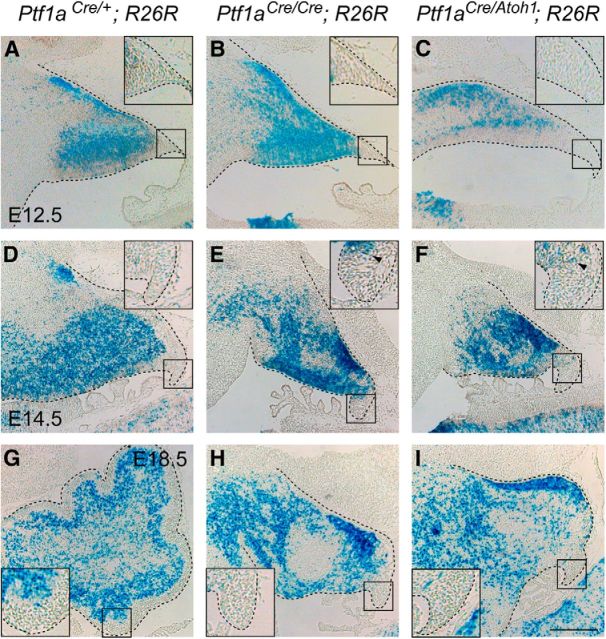
Figure 2.
Dynamics of Ptf1a lineage cells in the cerebellum during embryogenesis. A–I, X-gal-stained sagittal sections of the cerebellar primordia. Insets, High-magnification views of rectangular regions in A–I, respectively. Developmental stages and genotypes are indicated. Some stained cells found at the pial side in A are cells that are supposed to migrate out of the cerebellum (our unpublished data). Scale bar, 200 μm.

