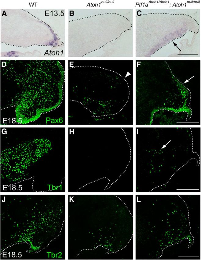
Figure 4.

Identity of cells produced in the cerebellum that expresses Atoh1 only in the VZ but not in the RL. A–C, Expression of all (endogenous + exogenous) Atoh1 transcripts in the cerebellum at E13.5. Black arrow in C indicates ectopic expression of Atoh1 in the VZ. D–L, Immunostaining with cell type-specific markers, such as Pax6 (D–F), Tbr1 (G–I), and Tbr2 (J–L), to the E18.5 cerebella. White dotted lines indicate the edge of the cerebellar primordium. All are sagittal sections. Top is dorsal, and left is rostral. WT, Wild-type. Scale bars, 200 μm.
