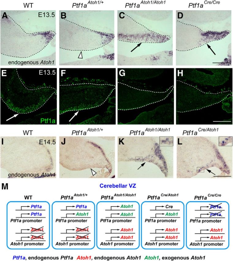
Figure 9.

Expression of the endogenous Atoh1 in the cerebellar primordium of the knock-in mice. A–H, Sagittal sections of cerebella of indicated genotypes at E13.5. Localization of endogenous Atoh1 (Atoh1 3′ UTR probes, in situ hybridization, A–D) and Ptf1a (immunofluorescence, E–H) is shown. I–L, Localization of endogenous Atoh1 transcripts in the cerebellar primordium of indicated genotypes at E14.5. Interestingly, ectopic expression of endogenous Atoh1 in the VZ was not detected in Ptf1aAtoh1/+ cerebella (J, white arrow) in contrast to those of Ptf1aCre/Atoh1 and Ptf1aAtoh1/Atoh1 (K, L, black arrows). Insets in K, L, High-magnification pictures of the boxed regions. Scale bars, 200 μm. M, Deduced schematic model for Ptf1a and Atoh1 transcription in cells of the cerebellar VZ. Genotypes are indicated. WT, Wild-type.
