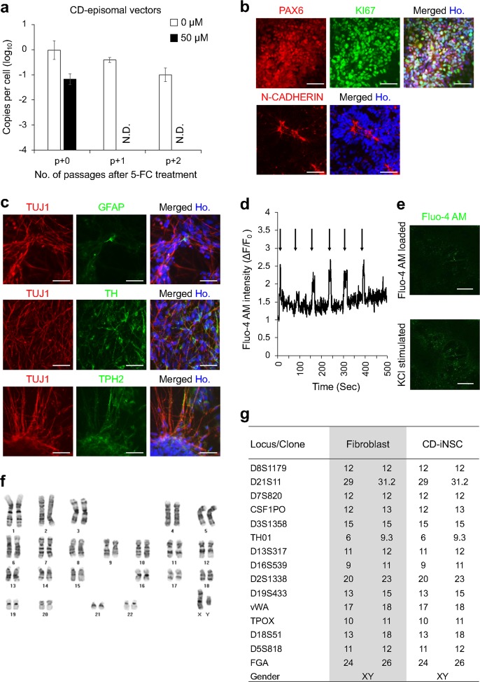
Fig. 5. Rapid elimination of residual CD episomal vectors in human-induced neural stem cells.
a qPCR analysis of total DNA in CD-iNSCs cultured with or without 5-FC was used to measure the copy numbers of residual CD episomal vectors. N.D. indicates not detected. b Immunocytochemistry of NSC markers that were expressed in the isolated EF-iNSCs. Scale bars represent 100 µm. c Images of immunocytochemical staining of neuronal and glial markers are shown to confirm the differentiation potential of isolated EF-iNSCs. d KCl-induced transient Ca2+ (real-time) in neurons at eight weeks post differentiation. The arrow represents KCl stimulation. e Representative images of Fluo-4 AM-loaded neurons pre and post KCl administration. Scale bars represent 200 µm. f G-banding analysis of EF-iNSCs showed normal human karyotypes. g STR analysis revealed that the CD-iNSCs were derived from the original CRL2097 fibroblasts

