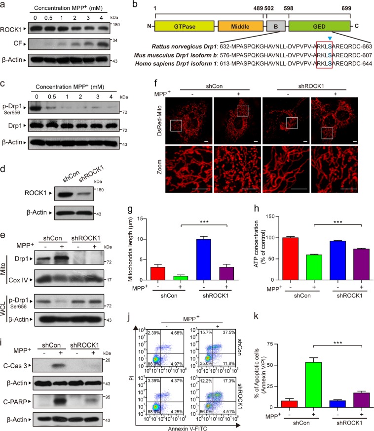
Fig. 3. ROCK1 activation is involved in MPP+-induced aberrant mitochondrial fission and apoptosis through the dephosphorylation/activation of Drp1.
a PC12 cells were treated with various concentrations of MPP+ (0, 0.5, 1, 2, 3, and 4 mM) for 24 h. The expression of ROCK1, CF of ROCK1, and p-Drp1 (Ser 656) in whole-cell lysates was determined by western blot analysis. CF represents the cleavage fragment of ROCK1. b The domain structure of Drp1. Highly conserved motifs in Drp1 isoforms were identified in Rattus, Mus musculus and Homo sapiens. c The expression of p-Drp1 (Ser 656) and total Drp1 in whole-cell lysates was determined by western blot analysis. d The stable expression of shCon or ROCK1 shRNA (shROCK1) in PC12 cells was confirmed by western blot analysis. Then the cells were treated with MPP+ (1 mM) alone or in combination with ROCK1 knockdown. e The expression of Drp1 in mitochondrial lysates (Mito) and p-Drp1 (Ser 656) in whole-cell lysates (WCL) was determined by western blot analysis. f Cells were transfected with a DsRed-Mito plasmid, and the mitochondria were viewed by confocal microscopy. Scale bars: 5 μm. g Mitochondrial length was quantified using Imaris software. h The ATP Determination Kit was used to determine the concentration of ATP. i The expression of C-Cas 3 and C-PARP in whole-cell lysates was determined by western blot analysis. j, k The apoptosis rate was measured by flow cytometry using annexin V-FITC/PI staining. The data are expressed as the means ± S.D. (n = 3). ***P < 0.001

