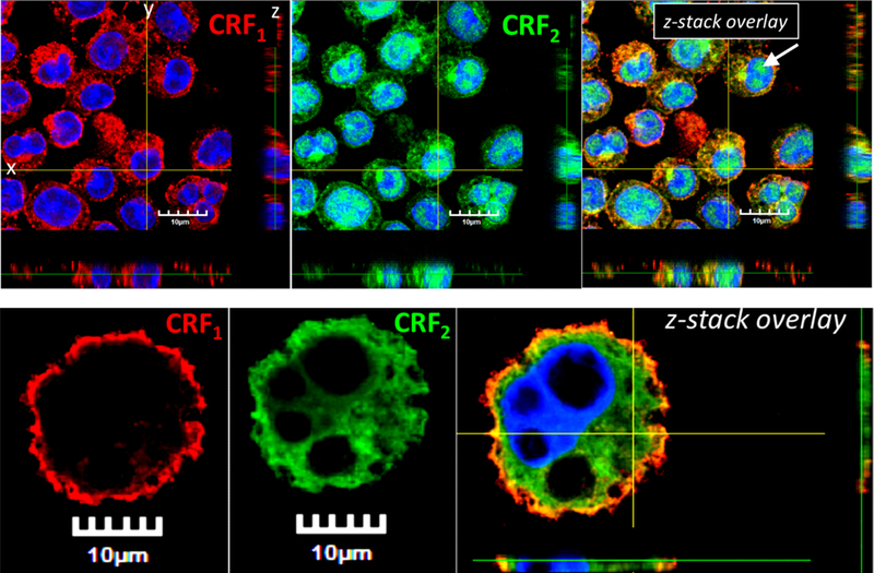
FIG 2. Localization of CRF1 and CRF2 in BMMCs and peritoneal mast cells.

CRF1 (Cy3 Red) and CRF2 (FITC Green) nuclei (DAPI blue). z-Stack overlay images show a single plane cross section through horizontal plane (XY), sagittal plane (YZ), and coronal plane (XZ). Panels A-C) murine BMMCs. Panels D-F) PMCs. White arrow indicates the nuclear staining pattern of CRF2 (Panel C).
