Figure 6.
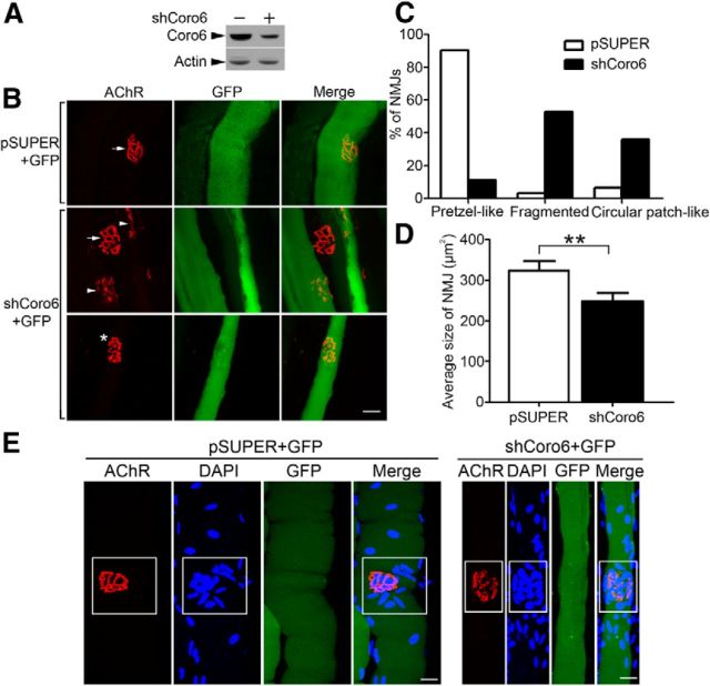
Coronin 6 knockdown perturbed AChR clustering in vivo. A, HEK-293T cells were cotransfected with Coronin 6 and pSUPER (−) or shCoro6 (+). Cell lysates were subjected to immunoblotting with antibodies against Coronin 6 or actin. B, The tibialis anterior muscles of adult mice were injected with 5 μg GFP and 30 μg pSUPER or shCoro6 followed by electroporation. Three weeks later, the muscles were stained with AlexaFluor-555-conjugated α-BTX to visualize AChR clusters. GFP signals indicate the transfected skeletal muscle fibers. Arrows indicate the normal pretzel-like structures of the NMJ. Impaired NMJ structures, such as fragmented (arrowhead) and circular patch-like structures (*), were observed in Coronin 6-knockdown fibers. C, Percentages of NMJs exhibiting pretzel-like, fragmented, and circular patch-like shapes (n = 31 from 4 mice injected with pSUPER; n = 36 from 4 mice injected with shCoro6). D, Quantification of the size of AChR clusters. **p < 0.01, shCoro6 versus pSUPER (Student's t test). E, Clustering of nuclei at the subsynaptic regions was unaltered in Coronin 6-silenced muscle. Nuclei and AChR clusters were visualized by DAPI (blue) and AlexaFluor-555-conjugated α-BTX staining (red), respectively. The subsynaptic regions are highlighted in rectangles. Scale bars, 20 μm.

