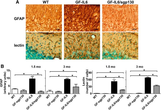
Figure 5.
IL-6 trans-signaling contributed to the development of gliosis in the cerebellum of GFAP-IL6 mice. A, Paraformaldehyde fixed, free-floating brain sections (30 μm) prepared from 1.5-month-old mice were processed for immunohistochemical detection of GFAP or lectin histochemistry. Scale bar, 25 μm. B, Staining in A and for similarly stained brain sections from 3-month-old mice were quantified with the NIH ImageJ analysis software. These analyses were performed on at least three blinded sections per brain and on a minimum of three brains per genotype. Values represent the mean ± SEM; *p ≤ 0.05.

