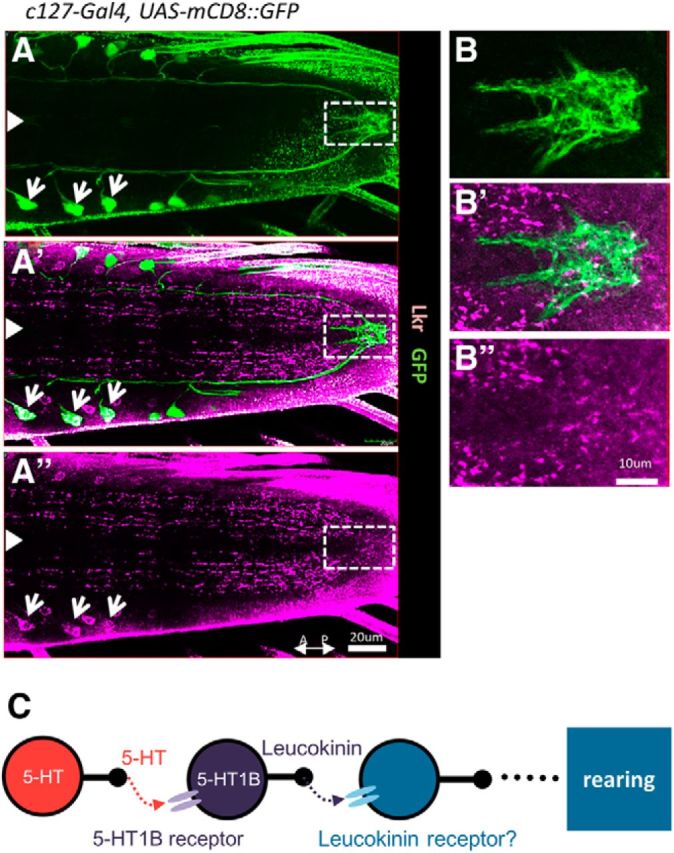
Figure 8.

Visualization of leucokinin receptors in the VNC. A–B″, Confocal images of larval VNCs stained for the leucokinin receptor (purple) and mCD8::GFP (expressed by c127-Gal4; green). Expression in ABLK cell bodies is indicated by white arrows. The region demarcated by white dotted rectangles in A–A″ is enlarged in B–B″ to show expression in the plexus near the axon terminals of ABLKs. White arrowheads indicate the midline. C, A model showing the signaling pathway of 5-HT involved in the regulation of rearing.
