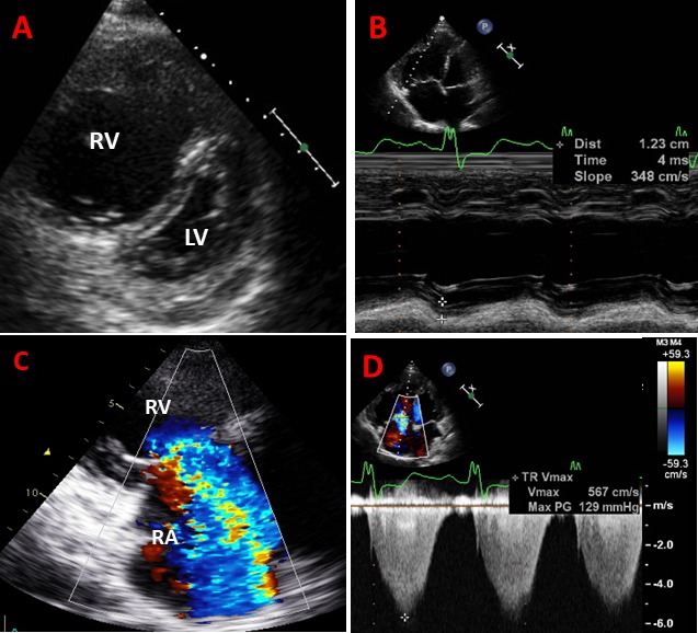
Figure 1.

Examples of images obtained from patient with pulmonary hypertension, right ventricular (RV) dilatation and dysfunction and torrential tricuspid regurgitation (TR). (A) Parasternal short-axis view shows severe dilatation of RV and flattened D-shape interventricular septum due to elevated RV pressure; (B) apical four-chamber view shows dilated RV with reduced tricuspid annular plane systolic excursion (TAPSE) of 1.2 cm; (C) torrential TR where colour jet occupies the entire right atrium (RA); and (D) peak velocity of tricuspid regurgitant jet by continuous-wave Doppler with an estimated systolic pulmonary artery pressure of 129 mm Hg. LV, left ventricle
