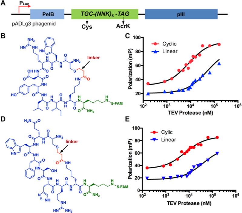
Figure 3.
Selected TEV protease-binding cyclic peptides and their Kd measurements. (A) A diagram to show the phagemid structure for the production of a phage-displayed 6-mer cyclic peptide library; (B) The structure of 5FAM-CycTev1. CycTev1, highlighted in blue and red, was selected from phage display; (C) Fluorescence polarization analysis of 5FAM-CycTev1 binding to TEV protease. Data for a linear counterpart of 5FAM-CycTev1 with no linker is also included; (D) The structure of 5FAM-CycTev2; (E) Fluorescence polarization analysis of 5FAM-CycTev2 binding to TEV protease. Data for a linear counterpart of 5FAM-CycTev2 with no linker is also included.

