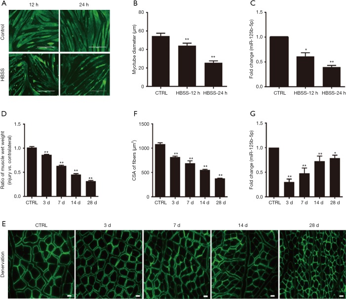
Figure 1.
Decreased expression of miR-125b-5p during muscle atrophy in vitro and in vivo. (A) Phenotypic changes of C2C12 myotubes following 12 or 24 h HBSS treatment (nutritional deprivation, fasting) or following no HBSS treatment (control, CTRL), as shown by MHC immunostaining (green). Scale bar, 400 µm. (B) Comparison in the average diameter of C2C12 myotubes, which had been treated as described in (A). **, P<0.01 vs. CTRL. (C) RT-qPCR data showing the expression level of miR-125b-5p in C2C12 myotubes, which had been treated as described in (A). *, P<0.05 and **, P<0.01 vs. CTRL. (D) Comparison in the wet weight ratio (the injured/contralateral side) of TA muscles of rats, which had been subjected to sciatic nerve cut before being killed at 3, 7, 14, and 28 days post-surgery. TA muscles of sham-operated rats served as CTRL. **, P<0.01 vs. CTRL. (E) Representative images of laminin immunostaining for measuring the cross-sectional area (CSA) of TA muscle fibers of rats, which had received treatments as described in (D). Scale bar, 20 µm. (F) Comparison in the CSA of rat TA muscle fibers, based on immunostaining data. **, P<0.01 vs. CTRL. (G) RT-qPCR analysis showing the decreased expression of miR-125b-5p in TA muscles of rats, which had received treatments as described in (D). *, P<0.05 and **, P<0.01 vs. CTRL. HBSS, Hank’s Balanced Salt Solution; TA, tibialis anterior; MHC, major histocompatibility complex.

