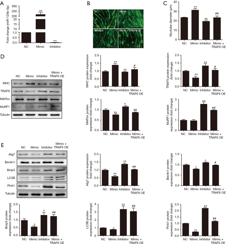
Figure 3.
Effect of miR-125b-5p on muscle atrophy in vitro. (A) RT-qPCR analysis showing the increased expression of miR-125b-5p in atrophic C2C12 myotubes after transfection with miR-125b-5p mimic and the decreased expression of miR-125b-5p after transfection with miR-125b-5p inhibitor, as compared to the expression of miR-125b-5p after vehicle transfection (negative control, NC). (B) Immunofluorescent staining showing phenotypic changes of atrophic C2C12 myotubes, which were transfected with miR-125b-5p mimic, miR-125b-5p inhibitor, or co-transfected with miR-125b-5p mimic and TRAF6 overexpression (OE) lentivirus, respectively. Scale bar, 400 µm. (C) Comparison of the average diameter of C2C12 myotubes after different treatments as mentioned in (B). (D) Western blot analysis of MHC, TRAF6, MAFbx, MuRF1 protein levels in atrophic C2C12 myotubes after different treatments as mentioned in (B). Tubulin served as a loading control. (E) Western blot analysis of the ALS related-protein levels in atrophic C2C12 myotubes after different treatments as mentioned in (B). Tubulin served as a loading control. In all bar graphs, *, P<0.05 and **, P<0.01 vs. NC; #, P<0.05 and ##, P<0.01 vs. transfection with miR-125b-5p mimic. TRAF6, tumour necrosis factor receptor adaptor protein 6; MHC, major histocompatibility complex; ALS, autophagy-lysosome system.

