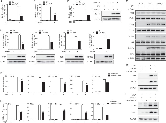
FIG 3.
Lnc-MxA restrains IFN-β transcription. (A and B) 293T cells were transfected with an IFN-β luciferase reporter along with an empty vector (EV) or an expression vector for lnc-MxA for 28 h. Then, the cells were infected with SeV at an MOI of 1 for 12 h (A) or stimulated with poly(I·C) (2 μg/ml) for 12 h (B). The cell lysates were harvested for a luciferase assay (n = 3; means and SD; *, P < 0.05; **, P < 0.01). (C) 293T cells were transfected with IFN-β luciferase reporter along with the indicated plasmids and either an empty vector or an expression vector for lnc-MxA for 28 h. The cell lysates were harvested for luciferase assay and immunoblotting analysis with the indicated antibodies (n = 3; means and SD; **, P < 0.01). (D) IFN-β mRNA expression was examined by RT-qPCR in 293T cells coexpressing lnc-MxA along with IRF3-5D. (E) 293T cells were transfected with an empty vector or an expression vector for lnc-MxA for 28 h. Then, the cells were infected with SeV or stimulated with poly(I·C). The cell lysates were collected and subjected to immunoblotting with the indicated antibodies. (F to I) A549-lnc-MxA and A549-ctrl cells were infected with SeV at an MOI of 0.1 (F and G) or influenza virus A/WSN/33 (H1N1) at an MOI of 0.1 (H and I) for 12 h. (F and H) Then, the total RNA and protein were prepared. The mRNA levels of the ISGs were determined by RT-qPCR (n = 3; means and SD; *, P < 0.05; **, P < 0.01). (G and I) The proteins were used for immunoblotting with antibodies against the indicated proteins.

