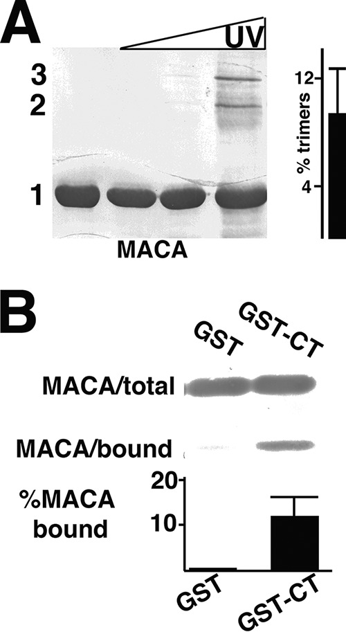
FIG 4.

Cross-linking and CT binding of WT MACA proteins. (A) Purified 50 μM WT HIV-1 MACA was UV cross-linked, electrophoretically separated, and detected as described in the legend to Fig. 2A. Monomer (1), dimer (2), and trimer (3) bands were determined by the mobilities of size standards run in parallel. The percentages of trimers relative to monomers are derived from four independent experiments. (B) WT MACA proteins were incubated with GST or GST-CT beads, and bound versus total MACA levels were detected as described in the legend to Fig. 2B. Relative percentages of bound versus total MACA protein levels were calculated from either two (GST) or five (GST-CT) independent experiments. The P value for the observed binding difference is 0.0083.
