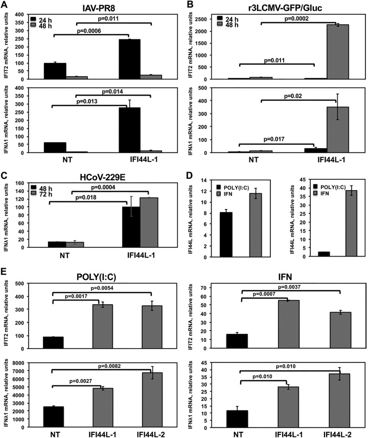
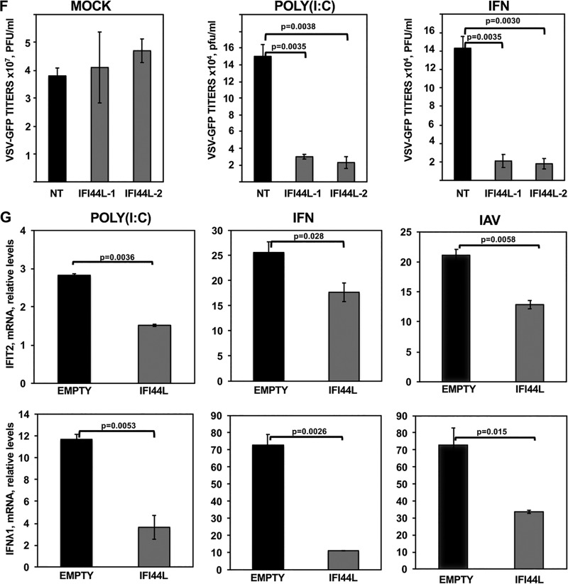
FIG 3.
IFI44L negatively modulates IFN responses. Human A549 (A and B) or Huh-7 (C) cells were transfected with the NT control siRNA and with an siRNA specific for IFI44L. At 36 hpt, A549 cells were infected with IAV PR8 strain (MOI, 0.1) (A) or with r3LCMV-GFP/Gluc (MOI, 0.1) (B) for 24 and 48 h, and Huh-7 cells were infected with HCoV-229E (MOI, 0.1) (C) for 48 and 72 h. Total RNAs were purified from infected cells, and expression levels of IFIT2 and IFN-λ1 mRNAs were evaluated by RT-qPCR and are represented as fold change values compared to values in mock-infected cells. (D, left panel, and E) Human A549 cells were transfected with two different siRNAs specific for IFI44L (E) or with the NT siRNA control (D, left panel, and E). (D, left panel, and E) At 36 hpt with the siRNAs, cells were transfected with poly (I·C) or treated with 250 U/ml of IFN-α. Expression levels of IFI44L (D), IFIT2 (E, top panels) and IFN-λ1 (E, bottom panels) were evaluated by RT-qPCR and are represented as fold change values compared to values in mock-treated cells. (F) After poly(I·C) transfection and IFN-α treatment, cells were infected with rVSV-GFP (MOI, 0.1) and virus titers in cell culture supernatants were determined at 24 hpi. (D, right panel, and G) Human 293T cells were transfected with pCAGGS-IFI44L-HA (G, IFI44L) or with an empty pCAGGS plasmid as a control (D, right panel, and G, EMPTY). (G) At 24 hpt, cells were transfected with poly(I·C) (left panels), treated with 1,000 U/ml of IFN-α (middle panels), or infected with IAV PR8 (MOI, 1) (right panels) for 24 h. Levels of IFI44L (D), IFIT2 (G, top panels), and IFN-λ1 (G, bottom panels) were evaluated by RT-qPCR. Bars represent SDs of results determined using duplicate wells. Three different experiments were performed, with similar results. P values determined by using a Student's t test are indicated for comparisons between NT-silenced cells and IFI44L-silenced cells.


