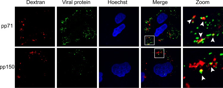
FIG 13.
HCMV tegument proteins coenter NT2 cells with the fluid-phase marker dextran. NT2 cells grown on coverslips were incubated with HCMV (AD169; MOI = 2) for 1 h at 4°C and then shifted to 37°C for 20 min in the presence of 5 μg/ml dextran (molecular weight, 10,000; dextran was conjugated to Texas Red). The cells were then fixed and processed for indirect immunofluorescence for the indicated viral tegument protein (green). Nuclei were stained with Hoechst (blue). Boxed areas are shown in magnification (zoom). White arrowheads indicate colocalization. Representative images of three independent biological replicates are shown.

