FIG 16.
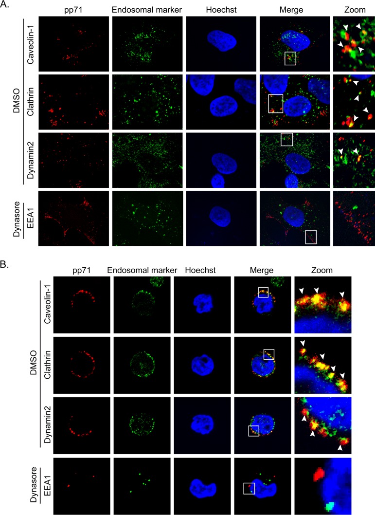
Endocytosis inhibitors impair HCMV virion internalization. (A) NT2 cells grown on coverslips were treated with dynasore (75 μM) or DMSO for 30 min and then infected with HCMV (AD169; MOI = 1) for 2 h at 37°C. The cells were then fixed and processed for indirect immunofluorescence for pp71 (red) and the indicated endosomal marker (green). Nuclei were stained with Hoechst (blue). Boxed areas are shown in magnification (zoom). White arrowheads indicate colocalization. Representative images of three independent biological replicates are shown. (B) THP-1 cells were treated with dynasore (75 μM) or DMSO for 30 min and then infected with HCMV (AD169; MOI = 10) for 4 h at 37°C. The cells were then fixed and processed for indirect immunofluorescence for pp71 (red) and the indicated endosomal marker (green). Nuclei were stained with Hoechst (blue). Boxed areas are shown in magnification (zoom). White arrowheads indicate colocalization. Representative images of three independent biological replicates are shown.

