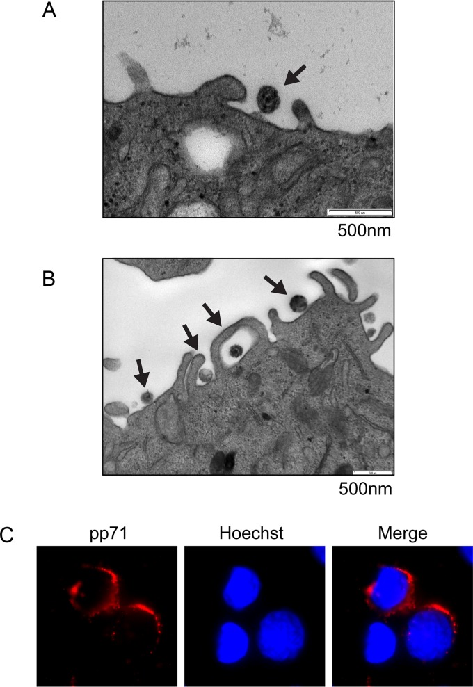
FIG 8.
Electron microscopy visualizes HCMV laboratory strain AD169 and clinical strain FIX entering THP-1 cells by macropinocytosis and endocytosis. (A) THP-1 cells were infected with HCMV (FIX; MOI = 10) for 2 h at 37°C and then processed for electron microscopy as described in Materials and Methods. The image is representative of six entry events detected in a single experiment. (B) THP-1 cells were infected with HCMV (AD169; MOI = 20) for 2 h at 37°C and then processed for electron microscopy as described in Materials and Methods. The image is representative of 57 entry events detected during five independent infections. (C) THP-1 cells were infected with HCMV (AD169; MOI = 20) for 2 h at 37°C and then processed for indirect immunofluorescence for pp71 (red). Nuclei were stained with Hoechst (blue). This nonconfocal image is representative of the images found in two independent biological replicates.

