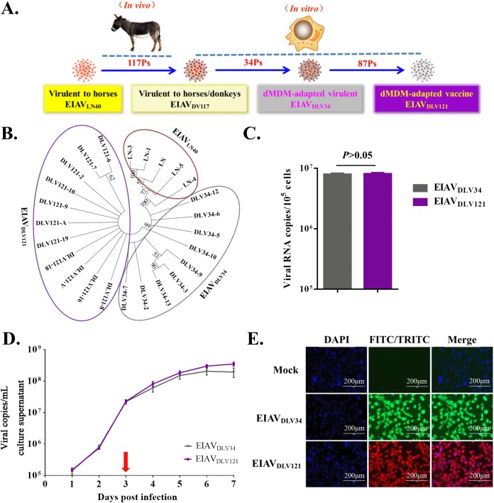
FIG 1.
Phylogenetic relationship and replication analyses of EIAVDLV34 and EIAVDLV121. (A) Process of passaging from the pathogenic virus strain to the attenuated vaccine strain. EIAVLN40, an EIAV strain isolated from Liaoning, China, has undergone 16 consecutive passages (Ps) in horses and is highly pathogenic to horses. After 117 consecutive passages in donkeys, this virus was transformed into strain EIAVDV117, which is highly virulent in both horses and donkeys. The virulence of EIAVDV117 gradually decreased after consecutive passages in dMDMs in vitro. In this study, EIAVDLV34 (the 34th generation, which is virulent for horses) and EIAVDLV121 (the 121th generation, which is avirulent and which induces protective immunity) were used for subsequent experiments. (B) Molecular phylogenetic analysis of the EIAVLN40, EIAVDLV34, and EIAVDLV121 sequences acquired through our previous studies. The evolutionary history was inferred by using a maximum likelihood method based on the general time-reversible model. Evolutionary analyses were conducted in MEGA (version 7) software (virus strains of different generations are represented by ellipses with different colors). (C) Comparison of the number of intracellular viral copies in the early phase of infection. The same infectious doses (5 × 10³ TCID50/well) of the pathogenic strain EIAVDLV34 and the attenuated EIAV strain EIAVDLV121 were used to infect eMDMs in 24-well microplates. At 3 h postinfection, the number of copies of intracellular EIAV RNA was measured by qPCR. (D) Comparison of the replication kinetics of EIAVDLV34 and EIAVDLV121 in eMDMs. Stocks of these two viruses with an equal TCID50 were used to infect eMDMs, as indicated. Viruses in the culture medium were quantified as viral RNA copy numbers at various time points up to 7 dpi. (E) Detection of eMDMs infected with EIAVDLV34 or EIAVDLV121 using an IFA. eMDMs were infected with EIAVDLV34 or EIAVDLV121, as indicated. The infected cells were then washed with PBS, fixed with 4% paraformaldehyde, and incubated with EIAV-positive horse serum. Cells infected with EIAV were visualized through incubation with FITC- or TRITC-conjugated rabbit anti-horse IgG and examined using fluorescence microscopy. DAPI, 4′,6-diamidino-2-phenylindole.

